Nikon NIS-Elements Microscope Imaging Software
Request a Quote or Demo
Never before has a microscope software package achieved such recognition for comprehensive device control with a suite of image analysis, visualization and archiving tools
Nikon’s NIS-Elements revolutionizes microscope imaging software for the microscopy market by combining automated intelligence to microscopes, cameras, components and peripherals with powerful analysis, visualization and archiving tools. Its intuitive interface simplifies workflow and speeds up image acquisition times while providing powerful features such as image stitching, object counting and volume views.
Multiple Options Scaled to Meet User Needs
The NIS-Elements software suite is composed of multiple distinct packages scaled to address specific application requirements. Each version delivers an intuitive interface to simplify workflow and provides powerful imaging tools. The software supports numerous image analysis and processing routines in conjunction with useful tools for storing and retrieving image data.
NIS-Elements Documentation (D)
NIS-Elements Documentation software provides a totally integrated solution for users of Nikon and other manufacturers’ accessories by delivering color documentation capabilities that include basic measuring and reporting.
NIS-Elements Documentation is tailored to facilitate image capture, object measurement and counting, databasing and report generation. Ease of use and simplification of menu options has been emphasized in the design of the package while NIS-Elements Advanced Research (AR) and Basic Research (BR) packages also display a similar look and feel.
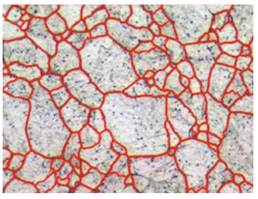
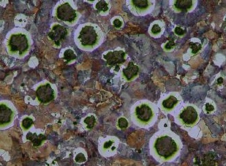
The combination of integrated automated intelligence, streamlined workflow, attention to detail and creative design makes NIS-Elements Documentation the perfect package for clinical and industrial applications such as tissue comparison, image archiving and reporting, particle analysis, defect analysis and fiber & textile material analysis.
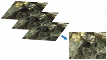
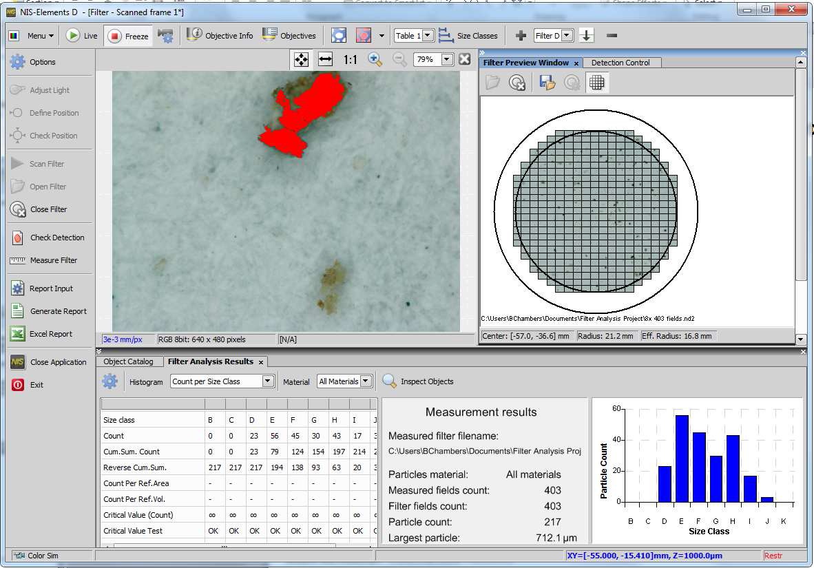
Core documentation functionality includes manual length and area measurements as well as large imaging stitching. Optional modules are available: database for streamlined image filing; retrieval and report generation for PDF based reports; Extended Depth of Focus (EDF), which creates an all-in-focus image from a series of Z-axis images; live compare for comparing a live image with a static image or overlay; Automatic Measurement for automated object counting using thresholding, feature restrictions and data export; and advanced macro builder for more complex custom programming.
NIS-Elements Basic Research (BR)
NIS-Elements BR is suited for standard research applications such as analysis and photodocumentation of fluorescent imaging. It features up to four dimensional acquisition capabilities and advanced device control capabilities.
While not as full featured as the AR package, BR offers access to advanced image capture, archiving and analysis solutions that are easy to use and provide maximum workflow.
BR handles multi-dimensional imaging with ease, with support for capture, display, peripheral device control, and data management and analysis of up to four dimensions (for example: X, Y, Z, Lambda (wavelength)). It also provides advanced image processing features, such as automated image count, intensity over time, database capabilities and report generation, and Extended Depth of Focus functions.
The system contributes to experiment efficiency with a database building feature developed to handle archiving, searching, and analysis of large numbers of multi-dimensional image files. Unified control of the entire imaging system offers significant benefits to users for cutting-edge research, such as live cell imaging.
NIS-Elements Advanced Research (AR)
NIS-Elements AR is optimized for advanced research applications. It features fully automated acquisition and device control through full six-dimensional image acquisition and analysis.
The software is designed to deliver power and maximize workflow. It handles multi-dimensional imaging flawlessly, with support for capture, display, peripheral device control and data management and analysis of up to six dimensions (X, Y, Z, Lambda (wavelength), T, multi stage points). It also offers sophisticated image processing and visualization features, such as a 5D viewer, automated object counting, intensity measurements over time, image stitching, deconvolution, databasing and Extended Depth of Focus functions.
The system contributes to experiment efficiency with a database building feature developed to handle archiving and searching of large numbers of multi-dimensional image files. Unified control of the entire imaging system offers significant benefits to users for cutting-edge research, such as live cell imaging.